
Visceral fat is fat stored around internal abdominal organs, distinct from the fat just under the skin. Most patients don't receive a direct visceral-fat number, so clinicians assess risk with proxies such as waist circumference and metabolic markers rather than appearance alone.
The popular advice to “lose belly fat” makes visceral fat sound like a cosmetic problem that can be solved with crunches, detox products, or a special supplement. That framing is misleading. Visceral fat is an invisible, metabolically active fat depot, and its clinical meaning depends on the person's overall health, not the shape of the abdomen or the number on a scale.
For adults considering physician-led obesity care, the useful questions are more specific: Where is the fat stored? What can BMI and waist circumference tell a clinician? When does imaging help, and when does it create false precision? And how should progress be followed when body weight doesn't tell the whole story?
Table of Contents
- What Visceral Fat Actually Is
- Visceral Fat Compared With Subcutaneous Fat
- Why BMI Alone Can Miss Visceral-Fat Risk
- How Clinicians Actually Assess Visceral-Fat Risk
- What the Research Actually Shows About Visceral Fat and Health Outcomes
- Why Diet and Exercise Affect Visceral Fat Differently Than People Expect
- When a Physician-Led Evaluation Is Worth Considering
- Common Questions About Visceral Fat
What Visceral Fat Actually Is
Visceral fat, also called visceral adipose tissue or VAT, is fat stored inside the abdominal cavity around organs. It differs from subcutaneous fat, which lies directly beneath the skin and can usually be pinched.
A simple analogy helps. Subcutaneous fat is like an outer layer of insulation beneath a coat. Visceral fat is more like an inner lining packed inside the coat, close to important equipment. The location changes how the tissue interacts with the rest of the body.
Visceral fat surrounds structures that include the liver and intestines. Enlarged visceral fat cells can contribute to the release of free fatty acids and inflammatory signals that affect liver metabolism and insulin sensitivity. That doesn't mean visceral fat alone determines a person's health. It means its location gives it different metabolic effects from fat stored under the skin, as described in this NIH-indexed review of visceral adipose tissue.
The distinction matters because body weight and BMI don't show fat distribution. Two adults can have the same BMI while carrying different amounts of abdominal fat. Conversely, a person whose BMI doesn't appear high can still have clinically meaningful visceral-fat risk.
The rest of the assessment therefore has three parts. First, the body's two main abdominal fat compartments need to be distinguished. Second, BMI and waist circumference need to be interpreted as screening tools with limits. Third, measurements need to be considered alongside blood pressure, glucose, cholesterol, liver markers, medical history, and treatment response.
Visceral fat is best understood as a metabolic-risk concept, not an aesthetic label. A physician can evaluate the broader pattern without treating a person's appearance as a diagnosis.
Visceral Fat Compared With Subcutaneous Fat
The two fat depots occupy different places and behave differently. Neither label can be assigned reliably from a mirror, a clothing size, or the feel of the abdomen alone.
Visceral vs Subcutaneous Fat at a Glance
| Feature | Visceral Fat | Subcutaneous Fat |
|---|---|---|
| Location | Deep inside the abdominal cavity around organs | Beneath the skin |
| Everyday visibility | Internal and not directly visible | May be visible or pinchable |
| Main clinical relevance | Metabolically active and associated with cardiometabolic risk | Provides insulation, energy storage, and physical cushioning |
| Direct measurement | More directly separated with CT or MRI | Easier to estimate with physical examination and body-composition methods |
| Common confusion | Mistaken for any protruding abdomen | Mistaken for the full amount of abdominal fat |
Subcutaneous fat is the tissue you notice when you think about body fat. It can sit on the abdomen, hips, thighs, arms, or other areas. Its presence doesn't automatically indicate poor health, and it isn't accurate to describe it as harmless in every circumstance. The key point is that it's located outside the abdominal cavity.
Visceral fat is deeper. CT research has shown that intra-abdominal fat includes omental, mesenteric, and retroperitoneal tissue, which helped researchers study fat distribution rather than relying only on body weight. Historical CT work also identified meaningful sex differences. Visceral-fat quantity can be up to two times higher in men than in premenopausal women, although distribution varies with age, hormonal status, ethnicity, and overall adiposity, according to this physiology review of visceral fat distribution.
Practical rule: A visible abdomen doesn't reveal how much fat is visceral, and a lean appearance doesn't prove that visceral-fat risk is absent.
A person with a larger body can have a relatively lower visceral-fat burden than another person with a smaller body. The reverse can also occur. That's why the words “stubborn belly fat” can obscure the medical question. The issue isn't whether fat looks resistant to exercise. The issue is whether the overall pattern suggests higher metabolic risk.
Physical activity still matters for general health and body composition. Resistance training is one component a clinician may discuss within an individualized plan, and this guide to resistance exercise for weight loss offers related educational context. It shouldn't be presented as a promise of spot reduction or as a way to diagnose visceral fat.
Why BMI Alone Can Miss Visceral-Fat Risk
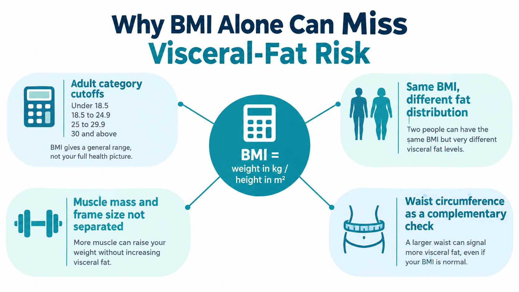
BMI is a screening measure based on weight relative to height. It can help organize an initial conversation, but it doesn't identify where fat is stored, distinguish fat from lean tissue, or measure visceral fat directly. The NIDDK explanation of weight assessment describes why BMI works best as one part of a broader assessment.
Where BMI can mislead
A muscular adult may have a higher BMI because of lean mass rather than a high visceral-fat burden. An older adult may have changes in muscle mass and height that make BMI less informative than it appears. Sex differences, hormonal status, ancestry, genetics, and overall adiposity also influence where fat accumulates.
The same BMI can therefore represent different body compositions and different metabolic profiles. BMI may be useful for screening, but it can't answer the question, “How much fat is around this person's organs?”
Research supports that limitation. In an NIH-indexed community study, each additional 100 cm² of visceral fat was associated with a 28% higher adjusted hazard of incident metabolic syndrome, after adjustment, and greater visceral-fat area was linked with higher cardiometabolic risk regardless of BMI. The association doesn't mean a scan result diagnoses metabolic syndrome or predicts an individual outcome. It shows why BMI shouldn't be used as the sole lens, as detailed in the community-based visceral-fat study.
Why population context matters
A single BMI interpretation can also miss population differences. Researchers and professional groups have discussed population-specific thresholds because cardiometabolic risk may emerge at different body sizes across groups. Those discussions don't justify applying an improvised cutoff to an individual. They reinforce the need to interpret BMI with waist circumference, laboratory findings, blood pressure, family history, and clinical history.
The BMI information from Empire Medical Wellness can provide general context, but BMI remains a starting point rather than a visceral-fat measurement. A thoughtful evaluation asks what BMI means in the context of the person in front of the clinician.
How Clinicians Actually Assess Visceral-Fat Risk
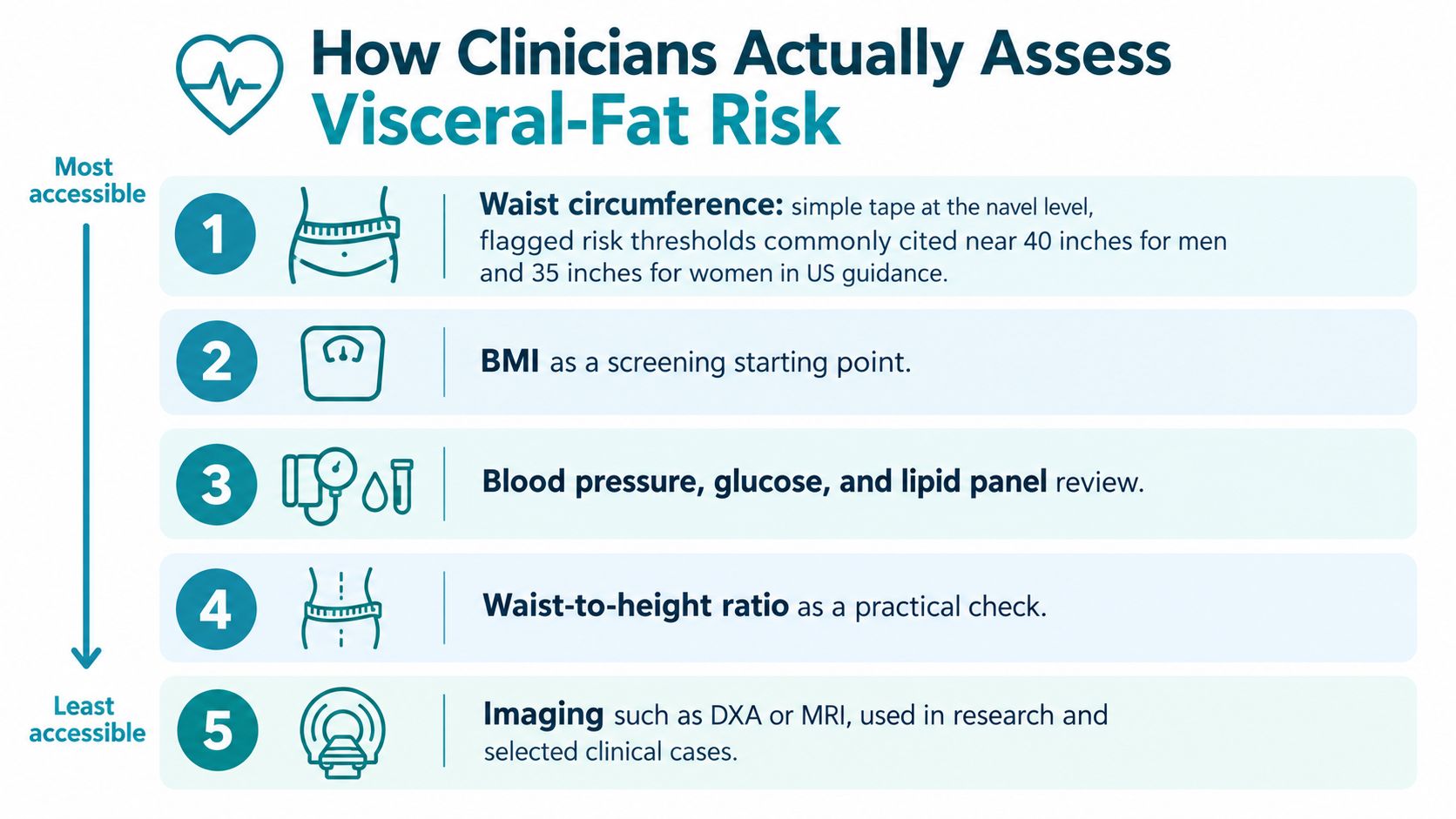
Most adults don't need a scan to begin a discussion about abdominal-fat risk. A clinician usually starts with accessible measures and then considers whether more detailed body-composition assessment would change the clinical picture.
Waist circumference as a proxy
Waist circumference estimates central adiposity, which includes both visceral and subcutaneous fat. It does not directly measure visceral-fat mass. A commonly used approach is to place a flexible tape horizontally around the abdomen and measure at the end of a gentle expiration. Professional guidance differs somewhat on the exact anatomical site, so consistency and documented technique matter. The IAS and ICCR waist-measurement consensus discusses these measurement considerations.
The NHLBI identifies a waist circumference above 40 inches for men and above 35 inches for women as an unhealthy range associated with increased health risk. These are screening reference points, not a standalone diagnosis, and they don't capture risk equally across sex, ancestry, age, or medical history, according to NHLBI guidance on overweight and obesity symptoms.
The broader metabolic picture
Blood pressure, fasting glucose or A1C, triglycerides, HDL cholesterol, liver markers, medication history, and family history can show health effects associated with visceral fat. None of those laboratory values directly measures visceral-fat volume. A normal result also doesn't prove that visceral fat is absent.
CT and MRI can separate visceral from subcutaneous fat more directly. DXA and other body-composition methods may provide useful information in selected settings, but results aren't interchangeable across technologies or protocols. CT isn't a routine self-monitoring tool because it involves radiation, and imaging access and cost also limit its role. Consumer bioimpedance scales can estimate body composition, but their visceral-fat scores shouldn't be treated as equivalent to CT or MRI.
For readers comparing available approaches, this overview of methods for measuring body composition can help explain why methods produce different kinds of information.
Measurement-limits checklist
- Waist circumference: Screens for central adiposity, but includes visceral and subcutaneous fat.
- BMI: Relates weight to height, but doesn't show fat distribution or separate fat from muscle.
- Blood pressure and laboratory markers: Describe cardiometabolic health, but don't measure visceral-fat volume.
- Consumer BIA: Produces an estimate that depends on the device and conditions, and isn't interchangeable with imaging.
- CT or MRI: Separates fat compartments more directly, but isn't routinely required for screening.
- Repeated measurements: Show direction over time only when the method, timing, and technique remain consistent.
A person preparing for a visit can review what to bring to a first obesity medicine appointment. The meaningful question usually isn't “What is the one correct visceral-fat score?” It's “What combination of measurements and health markers best explains this person's risk?”
What the Research Actually Shows About Visceral Fat and Health Outcomes
Higher visceral-fat measurements are associated with cardiometabolic problems, including insulin resistance, abnormal blood lipids, inflammatory activity, raised blood pressure, type 2 diabetes, and cardiovascular disease. The underlying pathways include portal delivery of free fatty acids to the liver, altered triglyceride processing, and inflammatory signaling. These are associations and mechanisms, not a guarantee of what will happen to any individual.
Mortality research illustrates why context matters. A 2022 systematic review of six cohorts found relative increases in all-cause mortality risk ranging from 11% to 98% across studies with higher abdominal VAT measurements. In one Pennington Biomedical Center cohort, each standard-deviation increase in VAT area, 70.1 cm², was associated with a 62% higher adjusted risk of death, with an adjusted hazard ratio of 1.62 and a 95% confidence interval of 1.07 to 2.47. These findings are reported in the systematic review of VAT and mortality.
The same evidence base also contains a less dramatic result. In the Multi-Ethnic Study of Atherosclerosis, each 50 cm² increase in VAT wasn't significantly associated with mortality. The adjusted hazard ratio was 1.02, with a 95% confidence interval of 0.93 to 1.12. Differences in age, population, follow-up, health status, measurement, and statistical adjustment can affect results.
Evidence boundary: Visceral fat is a meaningful risk marker, but no isolated measurement should be treated as a diagnosis, a guaranteed forecast, or a universal treatment target.
Emerging research has examined outcomes beyond diabetes and cardiovascular disease. A 2025 imaging study found that participants in the highest visceral-fat-area quartile had about twice the risk of incident diverticulitis compared with those in the lowest quartile, along with higher risks of complicated disease and recurrence requiring hospitalization. That finding is observational and doesn't prove that visceral fat directly caused those outcomes.
The practical conclusion is measured. Visceral fat deserves clinical attention, but a clinician needs to interpret it alongside age, sex, ethnicity, metabolic health, symptoms, family history, and the limitations of the measurement method. Adults can discuss related risk patterns through information on metabolic syndrome without turning a risk marker into a diagnosis.
Why Diet and Exercise Affect Visceral Fat Differently Than People Expect
Weight change and visceral-fat change are related, but they aren't identical. Two adults can lose the same amount of weight while experiencing different changes in visceral fat, subcutaneous fat, muscle, fitness, waist circumference, and metabolic markers.
A 2025 systematic review and meta-analysis found that, compared with exercise-only interventions, diet-only interventions didn't significantly change visceral-fat area, while exercise-only and combined diet-and-exercise interventions did. A separate 2025 meta-analysis of randomized trials found that prolonged exercise reduced abdominal visceral-fat area by 7.1% and subcutaneous-fat area by 9.1%, with higher exercise intensity associated with somewhat greater visceral-fat reduction, as reported in this 2025 exercise and visceral-fat meta-analysis.
Those findings don't mean every person will experience the same response, or that higher-intensity exercise is appropriate for everyone. They also don't support “spot reduction.” Abdominal exercises can strengthen muscles, but they haven't been established as a way to selectively remove fat from around abdominal organs. Detox products and supplements shouldn't be presented as proven visceral-fat treatments.
A more useful framework evaluates a repeatable activity plan and sustainable eating pattern against several outcomes:
- Waist trend: A consistent measurement can show changes in central size, although it still can't separate the two fat depots.
- Fitness and function: Improved strength, endurance, and daily activity can matter even when scale weight changes slowly.
- Metabolic markers: A1C, triglycerides, HDL cholesterol, blood pressure, and liver markers add clinical context.
- Muscle preservation: Resistance training can be considered as part of a broader plan, especially when maintaining function is a priority.
- Durability: A plan needs to fit a person's health, schedule, preferences, and treatment goals.
Educational material about coaching strategies for gym owners may help explain how adherence is supported in fitness settings, but it isn't a substitute for medical evaluation. Sleep, stress, nutrition, activity, and clinically appropriate obesity treatment can be discussed as connected parts of care, without promising selective visceral-fat loss.
When a Physician-Led Evaluation Is Worth Considering
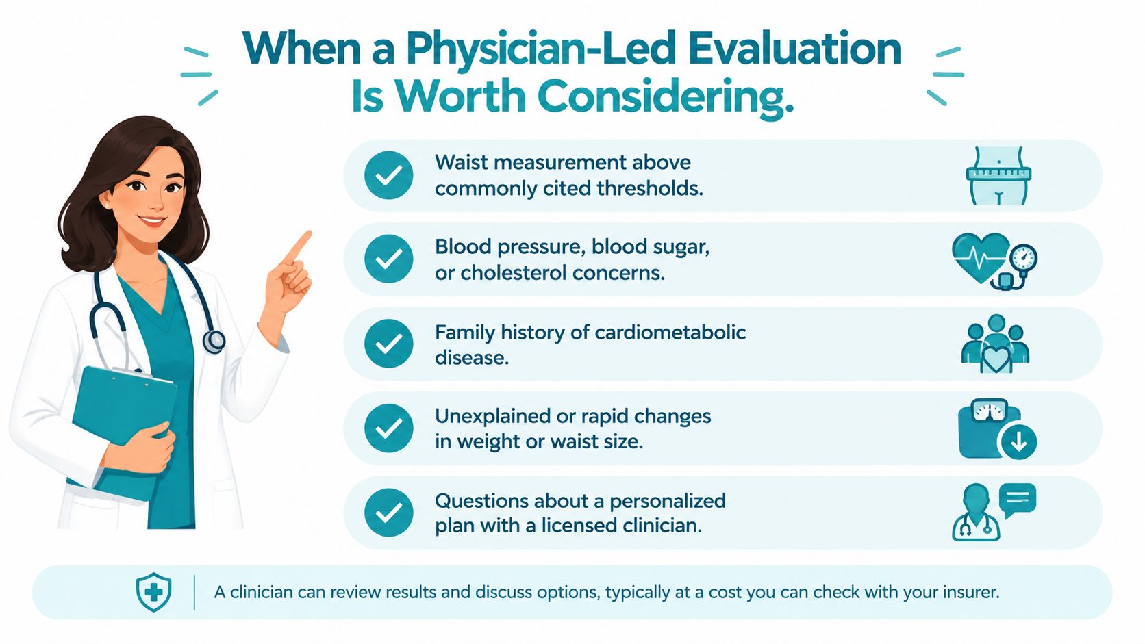
A physician-led obesity evaluation can be useful when visceral-fat concerns intersect with weight history, blood pressure, glucose, cholesterol, sleep, medications, menopause status, family history, or other health factors. The purpose isn't to chase a scan score. It's to understand the broader cardiometabolic picture and identify reasonable goals for monitoring and treatment.
A visit may include medical and metabolic assessment, review of prior weight-management efforts, individualized planning, discussion of anti-obesity medication when clinically appropriate, and follow-up based on treatment response. Medication decisions require a clinician's review of benefits, risks, contraindications, current medications, and medical history. No compounded medication, guaranteed result, or specific outcome should be assumed.
A blank follow-up worksheet
No patient measurements or three-month outcomes are supplied here. A consistent worksheet can help an adult and clinician compare change without inventing precision:
| Field | Entry |
|---|---|
| Date | ______________________________ |
| Measurement method | ______________________________ |
| Waist measurement and anatomical site | ______________________________ |
| BMI, if used | ______________________________ |
| Blood pressure | ______________________________ |
| A1C or fasting glucose | ______________________________ |
| Triglycerides and HDL cholesterol | ______________________________ |
| Relevant liver markers | ______________________________ |
| Activity, function, or sleep observations | ______________________________ |
| Clinician interpretation | ______________________________ |
| Follow-up plan | ______________________________ |
The method should remain consistent when change is being tracked. A waist measurement taken with the same technique may be more useful than switching repeatedly between a consumer scale, a scan, and a different tape-measure site. There's no universally valid visceral-fat cutoff across sex, age, ethnicity, BMI, or imaging modality. Longitudinal change plus outcomes such as waist circumference, A1C, triglycerides, blood pressure, or insulin-resistance markers is generally more actionable than an isolated scan value, as explained in this systematic review of visceral-fat thresholds.
Adults interested in physician-led obesity care can review weight-management services and decide whether a clinical conversation fits their needs. A physician can explain what is known, what remains uncertain, and which measurements would affect care.
Common Questions About Visceral Fat
Can a person with a normal BMI still have visceral-fat risk?
Yes. BMI doesn't show fat distribution, and research has linked greater visceral-fat area with cardiometabolic risk regardless of BMI. Waist circumference and metabolic markers can add information, but neither one independently diagnoses visceral fat.
Can ab exercises or a supplement target visceral fat?
No selective spot-reduction promise is supported here. Abdominal exercises may serve fitness or strengthening goals, but they shouldn't be marketed as a way to remove fat from around organs. Detox products and supplements also shouldn't replace evidence-based medical assessment.
Can a consumer BIA scale measure visceral fat reliably?
Consumer bioimpedance devices estimate body composition through device-specific algorithms. Their visceral-fat scores aren't interchangeable with CT or MRI, and a score shouldn't be treated as a diagnosis or a universal treatment target.
What should be recorded between visits?
A blank worksheet can include the date, the measurement method, waist measurement, relevant health markers, functional observations, and clinician interpretation. Repeating measurements under similar conditions helps show direction, but it doesn't convert a proxy into a direct visceral-fat measurement.
Adults who have questions about abdominal-fat risk can bring these observations to a qualified clinician rather than trying to interpret a single number alone. This article is educational and isn't personal medical advice.
Empire Medical Wellness offers physician-led obesity medicine evaluations that may include medical and metabolic assessment, individualized weight-management planning, and follow-up for cardiometabolic risk factors associated with visceral fat. Adults can visit Empire Medical Wellness to learn more about the practice and its obesity medicine care.